Bladder Cancer


The bladder is an organ located in the pelvic cavity that stores and discharges urine. Urine is produced by the kidneys, carried to the bladder by the ureters, and discharged from the bladder through the urethra. Bladder cancer accounts for approximately 90% of cancers of the urinary tract (renal pelvis, ureters, bladder, urethra).
According to the National Cancer Institute, the highest incidence of bladder cancer occurs in industrialized countries such as the United States, Canada, and France. Incidence is lowest in Asia and South America, where it is about 70% lower than in the United States.
Incidence of bladder cancer increases with age. People over the age of 70 develop the disease 2 to 3 times more often than those aged 55–69 and 15 to 20 times more often than those aged 30–54.
Bladder cancer is 2 to 3 times more common in men. In the United States, approximately 38,000 men and 15,000 women are diagnosed with the disease each year. Bladder cancer is the fourth most common type of cancer in men and the eighth most common type in women. The disease is more prevalent in Caucasians than in African Americans and Hispanics.
Types
Bladder cancer usually originates in the bladder lining, which consists of a mucous layer of surface cells that expand and deflate (transitional epithelial cells), smooth muscle, and a fibrous layer. Tumors are categorized as low-stage (superficial) or high-stage (muscle invasive).
In industrialized countries (e.g., the United States, Canada, France), more than 90% of cases originate in the transitional epithelial cells (called transitional cell carcinoma; TCC). In developing countries, 75% of cases are squamous cell carcinomas caused by Schistosoma haematobium (parasitic organism) infection. Rare types of bladder cancer include small cell carcinoma, carcinosarcoma, primary lymphoma, and sarcoma.


Cancer-causing agents (carcinogens) in the urine may lead to the development of bladder cancer. Cigarette smoking contributes to more than 50% of cases, and smoking cigars or pipes also increases the risk. Other risk factors include the following:
Age
Chronic bladder inflammation (recurrent urinary tract infections, urinary stones)
Consumption of Aristolochia fangchi (herb used in some weight-loss formulas)
Diet high in saturated fat
Exposure to second-hand smoke
External beam radiation
Family history of bladder cancer (several genetic risk factors identified)
Gender (male)
Infection with Schistosoma haematobium (parasite found in many developing countries)
Personal history of bladder cancer
Race (Caucasian)
Treatment with certain drugs (e.g., cyclophosfamide—used to treat cancer)
Exposure to carcinogens in the workplace also increases the risk for bladder cancer. Medical workers exposed during the preparation, storage, administration, or disposal of antineoplastic drugs (used in chemotherapy) are at increased risk. Occupational risk factors include recurrent and early exposure to hair dye, and exposure to dye containing aniline, a chemical used in medical and industrial dyes. Workers at increased risk include the following:
Hairdressers
Machinists
Printers
Painters
Truck drivers
Workers in rubber, chemical, textile, metal, and leather industries
Incidence and Prevalence
Causes and Risk Factors
Signs and Symptoms
The primary symptom of bladder cancer is blood in the urine (hematuria). Hematuria may be visible to the naked eye (gross) or visible only under a microscope (microscopic) and is usually painless. Other symptoms include frequent urination and pain upon urination (dysuria).
Diagnosis
Diagnosis of bladder cancer includes urological tests and imaging tests. A complete medical history is used to identify potential risk factors (e.g., smoking, exposure to dyes). Laboratory tests may include the following:
NMP22®BladderChek® (to detect elevated levels of tumor markers in the urine)
Urinalysis (to detect microscopic hematuria)
Urine cytology (to detect cancer cells by examining cells flushed from the bladder during urination)
Urine culture (to rule out urinary tract infection)
NMP22®BladderChek® is a urine test used to detect elevated levels of a nuclear matrix protein (called NMP22®). Bladder cancer increases levels of this protein in the urine, even during the early stages of the disease.
Results of this test, which is noninvasive and is performed in a physician’s office, are available during the patient’s office visit. Studies have shown that when used with cystoscopy, NMP22®BladderChek® may be more effective than other diagnostic tests (e.g., urine tests or cystoscopy alone).
Various imaging tests may also be performed. Intravenous pyelogram (IVP) is the standard imaging test for bladder cancer. In this procedure, a contrast agent (radiopaque dye) is administered through a vein (intravenously) and X-rays are taken as the dye moves through the urinary tract. IVP provides information about the structure and function of the kidneys, ureters, and bladder. Other imaging tests include CT scan, MRI scan, bone scan, and ultrasound.
If bladder cancer is suspected, cystoscopy and biopsy are performed. Local anesthesia is administered and a cystoscope (thin, telescope-like tube with a tiny camera attached) is inserted into the bladder through the urethra to allow the physician to detect abnormalities. In a biopsy, tissue samples are taken from the lesion(s) and examined for cancer cells. If the sample is positive, the cancer is staged using the tumor, node, and metastases (TNM) system.
Staging
Once the physician has determined that a tumor exists, the next step is to clarify the tumor’s status. Several questions will have to be answered: Is the tumor large or small? Does it lie within the lining of the bladder or has it extended into the surrounding tissue? Has the tumor spread to nearby lymph nodes? Has the tumor metastasized to distant sites within the body?
Fortunately, several systems have been developed to answer these questions. The most common of these — the TNM (tumor, node, metastasis) system — allows tumors to be classified, or “staged,” according to their overall characteristics. A biopsy is removed and sent to a histopathologist for examination under a microscope. The pathologist then assigns a stage and a grade to the tissue sample.
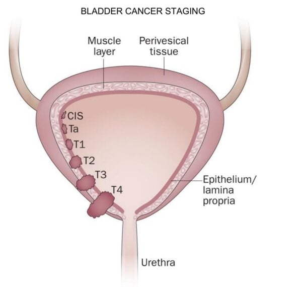
The stage refers to the physical location of the tumor within the bladder or, more specifically, the tumor’s depth of penetration. In general, the tumor stage is confined to one of two categories: (1) superficial, surface tumors, or (2) invasive, deep-spreading tumors. Superficial tumors affect only the bladder lining. They grow up and out from the lining tissue and extend into the bladder’s hollow cavity. Invasive tumors grow down into the deeper layers of bladder tissue, and they may involve surrounding muscle, fat, and/or nearby organs. Invasive tumors are more dangerous than superficial tumors since they are more likely to metastasize.
The grade is an estimate of the speed of tumor growth as suggested by cell features seen under a microscope. Most systems are based upon the degree of tumor cell anaplasia – that is, the loss of cellular “differentiation,” the distinguishing characteristics of a cell. The World Health Organization (WHO) grading system groups transitional cell carcinomas (TCCs) into three grades that correspond to well-, moderately, and poorly differentiated cells. The International Union Against Cancer (UICC) has devised a four-grade system that considers Grade 1 tumors to be well-differentiated, Grade 2 to be moderately differentiated and Grades 3 or 4 to be poorly differentiated. Both systems are widely used and can be summarized as follows:
Grade 1 (well-differentiated)
Grade 2 (moderately differentiated)
Grade 3 or Grade 4 (poorly differentiated)
There is a continuing debate about the classification of benign bladder lesions known as papillomas. The WHO defines papilloma as a single papillary (wart-like) growth with 8 or less cell layers in normal-looking surface tissue. By contrast, many pathologists and urologists classify papilloma as a Grade 1 TCC because of its tendency to recur and not to invade muscle.
There is a strong correlation between tumor stage and tumor grade. Nearly all superficial tumors are low grade; that is, they are Grade 1 tumors, with cells that are distinctly specialized and well-differentiated, whereas nearly all muscle-invasive tumors are high grade; that is, they are Grade 3 or 4 tumors, with cells that are nonspecialized and poorly differentiated. More importantly, there is a strong correlation between tumor stage and prognosis (the probable outcome of a disease), with superficial tumors having the most chance of a favorable result.
The latest TNM system for staging bladder cancer was developed by the UICC in 1997 (see Table 2).
T – Tumor
TX – Primary tumor cannot be evaluated
T0 – No primary tumor
Ta – Noninvasive papillary carcinoma
T1 – Tumor invades connective tissue under the epithelium (surface layer)
T2 – Tumor invades muscle
T2a – Superficial muscle affected (inner half)
T2b – Deep muscle affected (outer half)
T3 – Tumor invades perivesical (around the bladder) fatty tissue
T3a – microscopically
T3b – macroscopically (e.g., visible tumor mass on the outer bladder tissue)
T4 – Tumor invades any of the following: prostate, uterus, vagina, pelvic wall, abdominal wall
N – Regional Lymph Nodes
NX – Regional lymph nodes cannot be evaluated
N0 – No regional lymph node metastasis
N1 – Metastasis in a single lymph node < 2 cm in size
N2 – Metastasis in a single lymph node > 2 cm, but < 5 cm in size, or Multiple lymph nodes < 5 cm in size
N3 – Metastasis in a lymph node > 5 cm
M – Distant Metastasis
MX – Distant metastasis cannot be evaluated
M0 – No distant metastasis
M1 – Distant metastasis
Individuals with Grade 1, Stage 0 tumors usually do not need any additional workups for staging, because there is little risk of metastasis. By contrast, individuals with more advanced tumors, for example, Grade 2, Stage 2 tumors, require a routine staging assessment. Such an assessment should include basic blood work, chest X-ray, lower body imaging by either computed tomography (CT scan) or magnetic resonance imaging, and a bone scan.
Ta (papillary, noninvasive carcinoma)
“Ta” tumors are papillary (wart-like) in nature. They often look like pink cabbages, and they may be present in groups. Ta tumors are confined to the inner surface of the bladder wall and are distinguished from T1 tumors because they have not broken through the basement (supporting) membrane.
TIS (carcinoma in situ; flat, pre-invasive tumor)
Carcinoma in situ (CIS) of the transitional epithelium — otherwise known as TIS — is very rare. In the past, TIS tumors were associated with high death rates because they often were undiagnosed. Unlike papillary tumors, TIS tumors are flat. The cancerous cells in TIS tumors are pre-invasive (confined to the basement membrane). When detected in the urine by Pap staining, TIS cells appear anaplastic (lacking cellular differentiation – the distinguishing characteristics of a cell). In middle-aged men, TIS may resemble cystitis without hematuria. Accurate diagnosis depends upon biopsy of the mucosa in any patients with unexplained cystitis or sterile pyuria (no microorganisms are present but there is “pus-like” matter in the urine).
T1 (tumor invasion of connective tissue)
During clinical inspection, T1 tumors often look like Ta tumors. These cancers may appear as an isolated mass, or they may be present in groups. But the distinctive feature of the T1 tumor is that—although it has broken through the basement membrane into the connective tissue of the bladder-lining mucous membrane (lamina propria)—the stalk of the tumor has not invaded the muscle below. Some physicians believe that T1 tumors should not be considered “superficial TCC,” because they have the potential to be invasive and to progress. T1 tumors have a progression rate of roughly 30%. In T1 lesions of Grade 3 or Grade 4, nearly half of all tumors progress.
T2 (tumor invasion of muscle)
T2 tumors are characterized by the invasion of the muscle surrounding the bladder. If only the inner half of “superficial” muscle is affected (T2a tumor) and tumor cells are well-differentiated, the tumor may not have gained access to the lymphatic system. However, if the tumor has penetrated the outer half of “deep” muscle (T2b tumor) and cells are poorly differentiated, then the patient’s prognosis usually is worse.
T3 (tumor invasion of perivesical tissue)
When a tumor has broken through the surrounding muscle and begins to invade the perivesical tissue (fatty tissue around the bladder) or peritoneum (membrane lining the abdominal cavity) outside of the bladder, it is classified as a T3 tumor. If the process of invasion has just begun and only can be seen by microscopy, then the tumor is classified as T3a. However, if the tumor is visibly massed on the outer bladder tissue, then it is classified as T3b.
T4 (tumor invasion of surrounding organs)
If a tumor has progressed to invade nearby organs—such as the prostate (a male gland that surrounds the bladder neck and urethra and adds a secretion to the semen), uterus (womb), vagina (female reproductive canal), or walls of the abdomen or pelvis (hip bone)—it is classified as T4. T4 tumors are, by and large, inoperable, meaning they can/should not be surgically removed. They may cause painful symptoms, hematuria, frequent urination, and sleeplessness. In addition, the necrotic (dead) tissue within the bladder often becomes infected. Surgery may be performed not as a cure, but as a method to reduce suffering in patients with T4 tumors.
According to recent consensus decisions of the American Joint Committee on Cancer (AJCC), the stage groupings of bladder cancers are as follows:
Bladder cancer has a high rate of recurrence. Urine cytology and cystoscopy are performed every 3 months for 2 years, every 6 months for the next 2 years, and then yearly.
Bladder Cancer Follow-Up
Bladder Cancer Prognosis
Superficial bladder cancer has a 5-year survival rate of about 85%. Invasive bladder cancer has a less favorable prognosis. Approximately 5% of patients with metastasized bladder cancer live 2 years after diagnosis. Cases of recurrent bladder cancer indicate an aggressive tumor and a poor prognosis.
Bladder Cancer Prevention
Bladder cancer cannot be prevented. The best way to lower the risk is not to smoke. Studies have shown that drinking plenty of fluids daily also lowers the risk for bladder cancer.


